Transesophageal echocardiography is a method of ultrasound examination of the heart, in which the sensor is located not on the surface of the patient’s chest, but in the esophagus.
 Because the esophagus is behind the heart, the ultrasound beam does not need to travel through the chest wall and lungs to reach the heart. Because of this, the image of the organ is more clear and accurate.
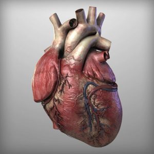
The researchers combined images from CT scans and transesophageal echocardiography to create a three-dimensional blueprint of the artificial heart. The internal organ was “printed” on a 3D printer https://en.wikipedia.org/wiki/3D_printing#Processes_and_printers. Scientists claim that the model turned out to be so accurate that it can be used for training surgical operations.
In the future, specialists will be able to create similar models for each individual patient and use them to diagnose people.